Imaging Services
The Imaging Center offers microPET, microCT, SPECT, MRI, bioluminescence and fluorescence imaging modalities and complementary in vitro/ex vivo services including cell-based assays, biodistribution, digital autoradiography and dosimetry. Companion PET tracer radiochemistry and radiolabeling services are available in-house and is supported by on-campus cyclotron facilities.
Technical and analytical support are available throughout the study process:
- Initial consultation
- Experimental design and optimization
- Imaging protocols and techniques
- Post-acquisition data analysis and interpretation
- Training and staff assistance
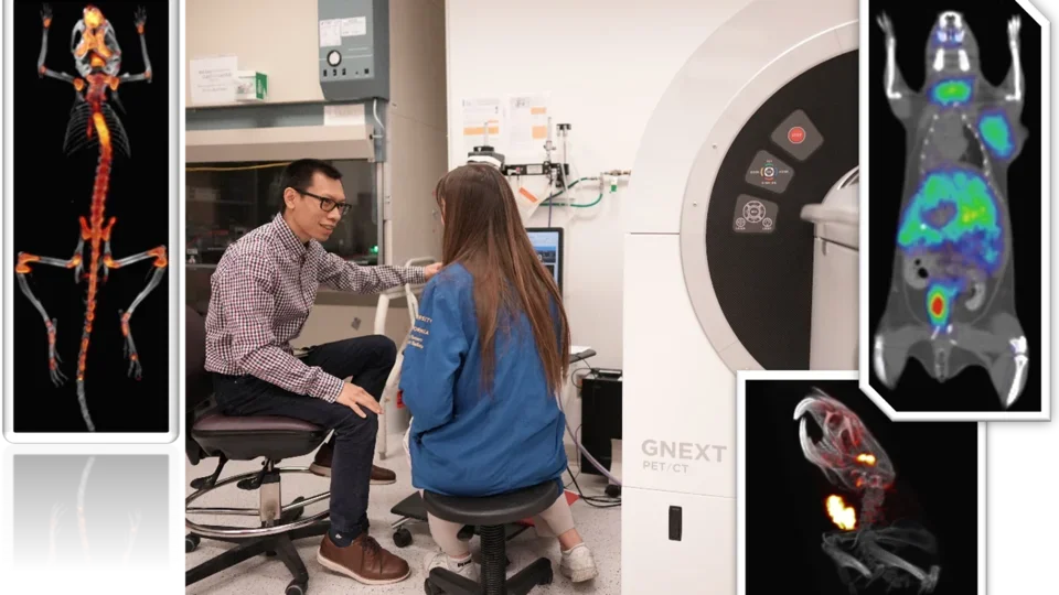
Positron emission tomography (PET) provides the means for imaging the rates of biologic processes in vivo. Imaging is accomplished through the integration of two technologies, the tracer kinetic assay method and computed tomography (CT). The tracer kinetic assay method employs a radiolabeled biologically active compound (tracer) and a mathematical model that describes the kinetics of the tracer as it participates in a biological process. The model permits the calculation of the rate of the process. The tissue tracer concentration measurement required by the tracer kinetic model is provided by the PET scanner, with the final result being a three-dimensional (3-D) image of the anatomic distribution of the biological process under study.
Excerpt from: Phelps, M.E., Positron Emission Tomography. In: Mazziotta, J. and Gilman, S., Eds., Clinical Brain Imaging: Principles and Applications, 1992, F.A. Davis Company, pp71-107
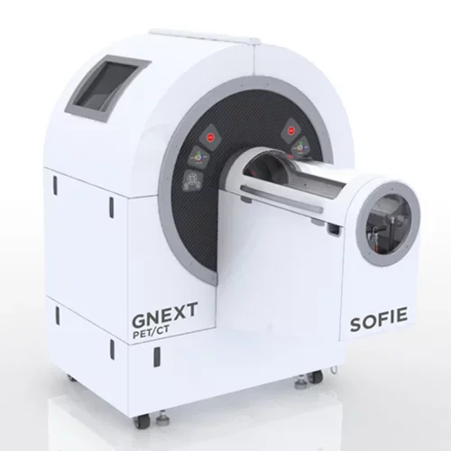
GNEXT PET/CT (Sofie Biosciences)
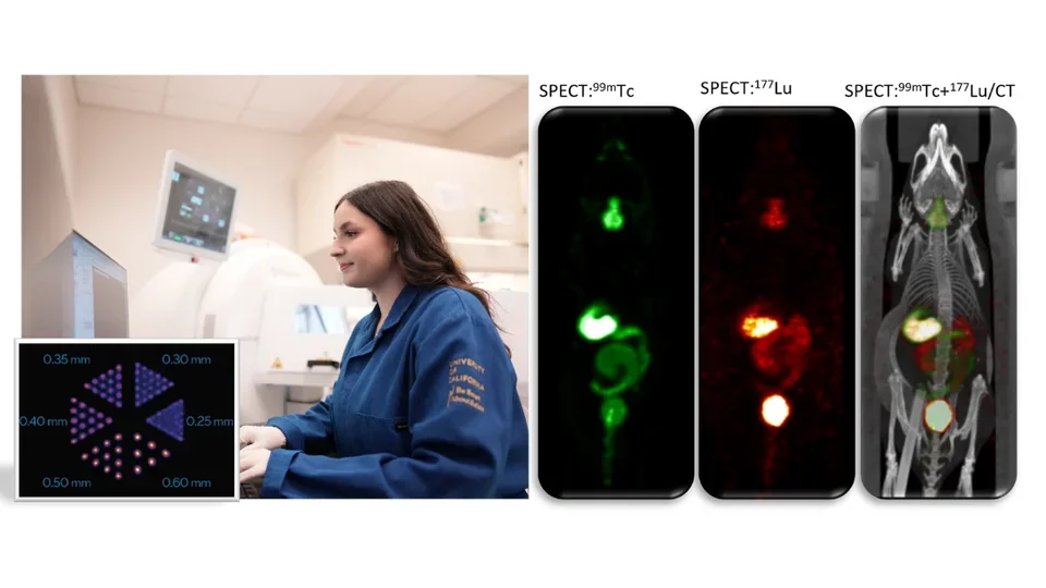
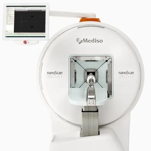
Single photon emission computed tomography (SPECT) imaging is a three-dimensional nuclear medicine imaging technique that enables real-time in vivo imaging to measure biodistribution of tracers, labeled novel compounds or cells. Our preclinical SPECT system (Mediso nanoScan SPECT/CT) combines an array of four gamma cameras on a gantry that rotate around the subject to provide spatial information on the distribution of the radionuclide within tissues. Common SPECT imaging applications include 99mTc sestamibi myocardial perfusion studies, 99mTc-HMPAO brain imaging to assess cerebral blood flow, 99mTc sestamibi parathyroid scans, 111In oxine-labeled white blood cell scans, 99mTc-methylene diphosphonate (MDP) bone scans, and biodistribution profiling of 177Lu- or 131I-labeled radioligand therapeutic agents.
nanoScan SPECT/CT (Mediso)
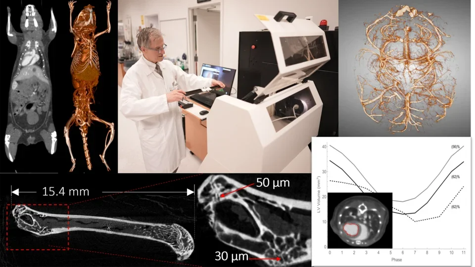
Computed tomography (CT), also known as computerized tomography or computed axial tomography, uses X-rays to produce detailed cross-sectional images of the body. Multiple X-ray projections from a single imaging session is reconstructed into a 3D image of the subject. With newer technologies available, the radiation doses imparted by these X-rays have been significantly reduced. As such, PET/CT imaging is standard in most preclinical and clinical studies to achieve accurate localization of PET measurements. Use of contrast such as iodine- or gold-based agents can enhance visualization of areas such as vasculature, abdominal tissues, and lymphatic tissues. Our current highest resolution in CT scan is 20 micron.
GNEXT (CT) (Sofie Biosciences)
HiCT (UCLA)
CrumpCAT (UCLA)
nanoScan SPECT/CT (Mediso)
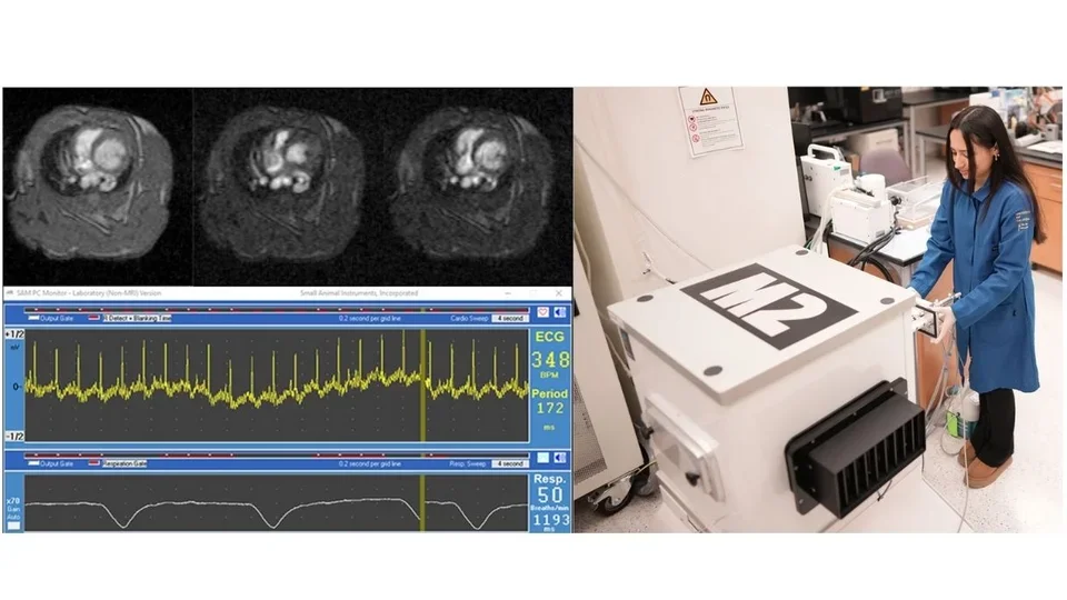
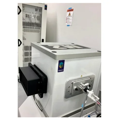
Magnetic resonance imaging (MRI), originally called nuclear magnetic resonance imaging (NMRI), is an imaging technique used to form pictures of the anatomy and the physiological processes of the body. MRI scanners use strong magnetic fields, magnetic field gradients, and radio waves to generate images of organs in the body. Because certain atomic nuclei are able to absorb radio frequency energy when placed in an external magnetic field, the resultant evolving spin polarization can induce a RF signal in a radio frequency coil and thereby be detected. Hydrogen atoms are naturally abundant in biological organisms, particularly in water and fat. For this reason, MRI scans essentially map the location of water and fat in the body. Pulses of radio waves excite the nuclear spin energy transition, and magnetic field gradients localize the polarization in space. By varying the parameters of the pulse sequence, different contrasts may be generated between tissues based on the relaxation properties of the hydrogen atoms therein. Compared to CT, MRI provides better contrast in images of soft-tissues, e.g. in the brain or abdomen. Our Aspect 1T M2 MRI has a spatial resolution of 156 micron, and can perform ECG-gated imaging of cardiovascular and respiratory systems.
M2 MRI for mice and rats (Aspect Imaging)
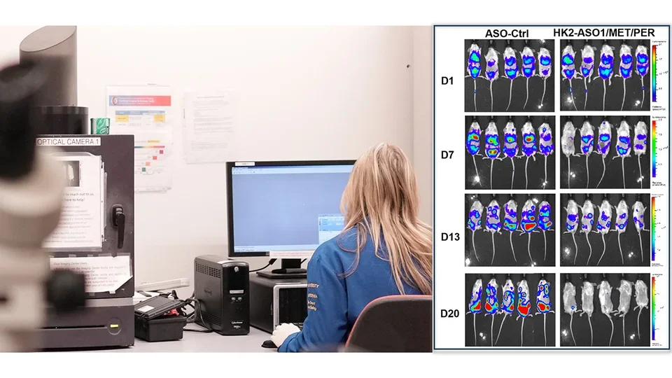
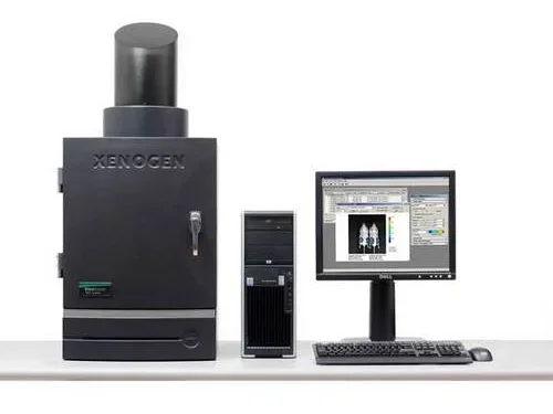
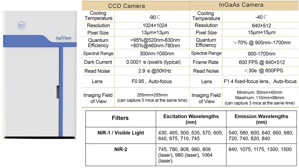
Optical imaging is a non-invasive technology to visualize and measure light produced through various means. In vivo optical imaging generally implies bioluminescence, light produced by enzymatic reactions, or fluorescence, light emitted from substances that have absorbed light. Cerenkov radiation from radiolabeled probes can also be detected by optical scanners. Optical reporter systems are extensively used by biomedical researchers for monitoring disease progression, cell trafficking, drug screening and therapeutic response. This assay can be further engineered to interrogate the functional states of cells using activatable reporter genes or probes.
IVIS Lumina II (Perkin Elmer)
Aniview Phoenix 100 (BioLight)
Plasmids for fluorescent proteins may be available at: AddGene
Managed by the UCLA Division of Laboratory Animal Medicine (DLAM)1, a dedicated vivarium adjacent to the Imaging Center offers certified veterinary, diagnostic laboratory, husbandry and procurement services. DLAM also provides preclinical toxicology and safety pharmacology studies under Good Laboratory Practice (GLP) compliance. An efficient and cost-effective mechanism has been established to fulfill Federal Drug Administration (FDA) regulatory requirements for translating promising PET imaging probes from preclinical research into human clinical trials2. The Imaging Center and DLAM are also developing the means to routinely offer various animal models for non-invasive, longitudinal imaging studies.
- Mosessian, S., et al. INDs for PET molecular imaging probes-approach by an academic institution. Molecular imaging and biology : MIB : the official publication of the Academy of Molecular Imaging 16, 441-448 (2014).
The Imaging Center or trained users can perform quantitative ex vivo biodistribution studies in small animals by cut-and-count tissue radioactivity measurements. Clinical translation of novel PET tracers or radiotherapeutic agents requires assessment of radiation dose exposure to human tissue. Dosimetry from mouse biodistribution studies is valuable for translation to human imaging and cancer radioligand therapy.
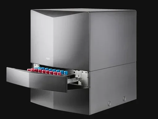
AMG Automatic Gamma Counter with Internal Scale (HiDex)
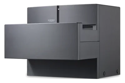
300 SL Liquid Scintilation Counter (HiDex)

Digital whole-body autoradiography (DWBA) of 18F-FDG biodistribution in a C57BL6 mouse.
Autoradiography permits hi-resolution assessment of radiolabeled molecules biodistribution. In particular, this technique is routinely used to complement PET biodistribution studies to further define the specific localization of PET probes. Services include whole body and tissue sectioning and phosphor imaging systems.
Leica CM3600 XP cryomacrotome – whole body sectioning
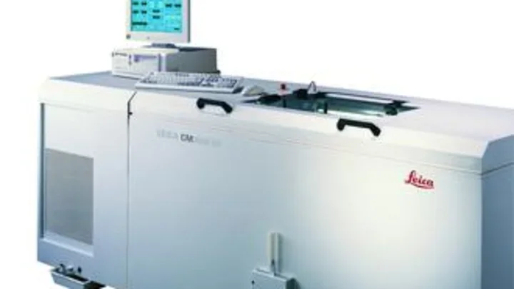
Leica CM3050 cryomicrotome – tissue sectioning
FUJIFILM BAS-500 phosphor imaging system