Imaging Services

The Imaging Center offers microPET, microCT, SPECT, MRI, bioluminescence and fluorescence imaging modalities and complementary in vitro/ex vivo services including cell-based assays, biodistribution, digital autoradiography and dosimetry. Companion PET tracer radiochemistry and radiolabeling services are available in-house and is supported by on-campus cyclotron facilities.

Technical and analytical support are available throughout the study process:

  • Initial consultation
  • Experimental design and optimization
  • Imaging protocols and techniques
  • Post-acquisition data analysis and interpretation
  • Training and staff assistance
Positron Emission Tomography (PET)
PET

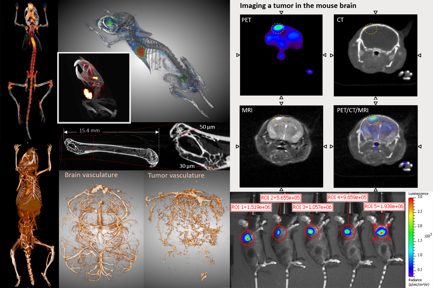
Positron emission tomography (PET) provides the means for imaging the rates of biologic processes in vivo. Imaging is accomplished through the integration of two technologies, the tracer kinetic assay method and computed tomography (CT). The tracer kinetic assay method employs a radiolabeled biologically active compound (tracer) and a mathematical model that describes the kinetics of the tracer as it participates in a biological process. The model permits the calculation of the rate of the process. The tissue tracer concentration measurement required by the tracer kinetic model is provided by the PET scanner, with the final result being a three-dimensional (3-D) image of the anatomic distribution of the biological process under study.

Excerpt from: Phelps, M.E., Positron Emission Tomography. In: Mazziotta, J. and Gilman, S., Eds., Clinical Brain Imaging: Principles and Applications, 1992, F.A. Davis Company, pp71-107

GNEXT PET/CT (Sofie Biosciences)

×

Genisys 8 PET/CT (Sofie Biosciences)

×
Single Photon Emission Computed Tomography (SPECT)
SPECT

Single photon emission computed tomography (SPECT) imaging is a three-dimensional nuclear medicine imaging technique that enables real-time in vivo imaging to measure biodistribution of tracers, labeled novel compounds or cells. Our preclinical SPECT system (Mediso nanoScan SPECT/CT) combines an array of four gamma cameras on a gantry that rotate around the subject to provide spatial information on the distribution of the radionuclide within tissues. Common SPECT imaging applications include 99mTc sestamibi myocardial perfusion studies, 99mTc-HMPAO brain imaging to assess cerebral blood flow, 99mTc sestamibi parathyroid scans, 111In oxine-labeled white blood cell scans, 99mTc-methylene diphosphonate (MDP) bone scans, and biodistribution profiling of 177Lu- or 131I-labeled radioligand therapeutic agents.

Computed Tomography (CT)
CT

Computed tomography (CT), also known as computerized tomography or computed axial tomography, uses X-rays to produce detailed cross-sectional images of the body. Multiple X-ray projections from a single imaging session is reconstructed into a 3D image of the subject. With newer technologies available, the radiation doses imparted by these X-rays have been significantly reduced. As such, PET/CT imaging is standard in most preclinical and clinical studies to achieve accurate localization of PET measurements. Use of contrast such as iodine- or gold-based agents can enhance visualization of areas such as vasculature, abdominal tissues, and lymphatic tissues. Our current highest resolution in CT scan is 20 micron.

CrumpCAT (UCLA)

×

HiCT (UCLA)

×

GNEXT (CT) (Sofie Biosciences)

×

Genisys 8 (CT) (Sofie Biosciences)

×
Magnetic Resonance Imaging (MRI)
MRI

Magnetic resonance imaging (MRI), originally called nuclear magnetic resonance imaging (NMRI), is an imaging technique used to form pictures of the anatomy and the physiological processes of the body. MRI scanners use strong magnetic fields, magnetic field gradients, and radio waves to generate images of organs in the body. Because certain atomic nuclei are able to absorb radio frequency energy when placed in an external magnetic field, the resultant evolving spin polarization can induce a RF signal in a radio frequency coil and thereby be detected. Hydrogen atoms are naturally abundant in biological organisms, particularly in water and fat. For this reason, MRI scans essentially map the location of water and fat in the body. Pulses of radio waves excite the nuclear spin energy transition, and magnetic field gradients localize the polarization in space. By varying the parameters of the pulse sequence, different contrasts may be generated between tissues based on the relaxation properties of the hydrogen atoms therein. Compared to CT, MRI provides better contrast in images of soft-tissues, e.g. in the brain or abdomen. Our Aspect 1T M2 MRI has a spatial resolution of 156 micron, and can perform ECG-gated imaging of cardiovascular and respiratory systems.

Optical (bioluminescence and fluorescence)
optical

Optical imaging is a non-invasive technology to visualize and measure light produced through various means. In vivo optical imaging generally implies bioluminescence, light produced by enzymatic reactions, or fluorescence, light emitted from substances that have absorbed light. Cerenkov radiation from radiolabeled probes can also be detected by optical scanners. Optical reporter systems are extensively used by biomedical researchers for monitoring disease progression, cell trafficking, drug screening and therapeutic response. This assay can be further engineered to interrogate the functional states of cells using activatable reporter genes or probes.

IVIS Lumina II (Perkin Elmer)

×

Maestro (CRi)

×
List of Optical Reagents
Modality Reporter system Size (kDa) Substrate Kinetics Secreted reporter gene? Peak Emission (nm)
Bioluminescence cypridina luciferase 61 cypridina luciferin glow yes 465
Bioluminescence dinoflagellate luciferase 40 dinoflagellate luciferin flash no 474
Bioluminescence gaussia luciferase 20 coelenterazine flash yes (non-secreted variant available) 480
Bioluminescence bacteria luciferase (luxABCDE - multiple enzyme system) varying endogenously produced - enzyme encoded by lux operon glow no 490
Bioluminescence renilla luciferase and variants 36 coelenterazine flash no 475-535 (variants)
Bioluminescence click beetle green 64 D-luciferin glow no 540
Bioluminescence firefly luciferase and variants 61 D-luciferin glow no 560
Bioluminescence click beetle red 64 D-luciferin glow no 620
Fluorescence visit this link for detailed information: http://www.fpvis.org

Plasmids may be available at: http://www.addgene.org/

Animal Models, Toxicology and FDA Studies
animal

Managed by the UCLA Division of Laboratory Animal Medicine (DLAM)1, a dedicated vivarium adjacent to the Imaging Center offers certified veterinary, diagnostic laboratory, husbandry and procurement services. DLAM also provides preclinical toxicology and safety pharmacology studies under Good Laboratory Practice (GLP) compliance. An efficient and cost-effective mechanism has been established to fulfill Federal Drug Administration (FDA) regulatory requirements for translating promising PET imaging probes from preclinical research into human clinical trials2. The Imaging Center and DLAM are also developing the means to routinely offer various animal models for non-invasive, longitudinal imaging studies.

  1. DLAM (Dr. Marcelo Couto): https://portal.dlam2.ucla.edu/Pages/Default.aspx
  2. Mosessian, S., et al. INDs for PET molecular imaging probes-approach by an academic institution. Molecular imaging and biology : MIB : the official publication of the Academy of Molecular Imaging 16, 441-448 (2014).
Biodistribution and Dosimetry
biodistribution

Biodistribution of 18F-D-FAC in C57BL6 mice for dosimetry calculations. Radu CG et al. Nat Med 2008

The Imaging Center or trained users can perform quantitative biodistribution studies in small animals by PET imaging or ex vivo tissue radioactivity measurements. Clinical translation of novel PET tracers require assessment of radiation dose exposure to human tissue. The Imaging Center is developing the service to offer PET probe dosimetry from mice biodistribution studies for translation to human imaging.

Wizard 3" automatic gamma counter (Perkin Elmer)

×

Inveon PET (Siemens)

×

Focus 220 PET (Concorde Microsystems)

×
Digital Autoradiography
autorad

Digital whole-body autoradiography (DWBA) of 18F-FDG biodistribution in a C57BL6 mouse.

Autoradiography permits hi-resolution assessment of radiolabeled molecules biodistribution. In particular, this technique is routinely used to complement PET biodistribution studies to further define the specific localization of PET probes. Services include whole body and tissue sectioning and phosphor imaging systems.

Leica CM3600 XP cryomacrotome – whole body sectioning

×

Leica CM3050 cryomicrotome – tissue sectioning

×

FUJIFILM BAS-500 phosphor imaging system

×
In Vitro Assays
invitro

In vitro technologies and assays of PET probe metabolism.
Vu NT et. al. JNM 2011
Wang J et.al. JNM 2013
Clark PM et al. PNAS 2014

A radiation- and BSL2-certified set-up in the Imaging Center is available for routine cell culture, sterile preparation and performing in vitro assays such as screening novel PET probes, optical reporter systems or therapeutics prior to in vivo imaging.

Cell culture set-up

×

Wizard 3" automatic gamma counter (Perkin Elmer)

×

Radiochemistry Services

The Radiochemistry center operates a cyclotron and a state-of-the-art radiochemistry laboratory to produce a wide variety of PET imaging tracers, and has access to additional compounds used in clinical practice and trials at UCLA via collaboration with the Ahmanson Translational Theranostics Division.

Though our primary focus is on 18F-labeled radiopharmaceuticals for PET imaging, we also work with other PET isotopes (64Cu, 68Ga, 124I, 89Zr) and therapeutic isotopes (177Lu) and have experience labeling peptides, proteins, and nanoparticles.

PET Tracer Production
PET tracers

By leveraging cutting-edge radiosynthesis technologies, we are able to offer a wide range of tracers on a per-batch basis. Generally, production must be requested at least 2 weeks in advance to allow sufficient time for ordering any needed materials and for coordinating any associated imaging studies, as applicable.

Standard analytical test results are available with all batches (e.g. radiochemical identity and purity). Please contact us if specialized tests (e.g. molar activity) are needed.

Available PET Tracers

The Radiochemistry Center is fully-equipped for production of 18F-labeled tracers. By leveraging cutting-edge technologies for PET tracer production, we are able to offer a wide range of tracers. Generally, production must be requested at least 2 weeks in advance to allow sufficient time for ordering any needed materials and for coordinating any associated imaging studies, if applicable.

PET Probes Type Biological Application
18F-FDG glucose glycolysis (cancer biology, neuroscience, etc)
18F-NaF bone appetite crystals bone metabolism; cancer biology (e.g. prostate cancer bone metastasis)
18F-FLT thymidine (nucleoside) proliferation; nucleoside metabolism (e.g. cancer biology, immunology)
18F-FHBG nucleoside reporter gene imaging
18F-D-FAC (and analogs) deoxycytidine (nucleoside) immunology (e.g. adaptive immune response; immunotherapies)
cancer biology (e.g. treatment stratification of nucleoside analog prodrugs)
18F-L-FMAU (and analogs) nucleoside reporter gene imaging
18F-D-FEAU nucleoside reporter gene imaging (e.g. cell trafficking and cellular function)
124I-FIAU nucleoside reporter gene imaging (e.g. cell trafficking and cellular function)
18F-Fallypride D2 receptor ligand neuroscience
18F-FDHT testosterone cancer biology (prostate cancer)
18F-AraG guanosine (nucleoside) immunology; cancer biology
18F-FDOPA amino acid (phenylalanine) dopamine synthesis: neuroscience & brain tumors
18F-FDDNP amyloid ligand neuroscience: Alzheimer's & other amyloid disorders
11C-acetate acetate cancer biology (prostate cancer)
fatty acid synthesis
13N-ammonia ammonia blood flow at the capillary level (perfusion)
11C-L-glutamine glutamine cancer biology; glutamine metabolism
18F-clofarabine (18F-CFA) deoxycytidine (nucleoside) immunology (e.g. adaptive immune response; immunotherapies)
cancer biology (e.g. treatment stratification of nucleoside analog prodrugs)
18F-flumazenil (18F-FMZ) benzodiazepene receptor ligand neuroscience
18F-florbetaben (18F-FBB) amyloid ligand neuroscience: Alzheimer's & other amyloid disorders
18F-FPEB metabotropic glutamate receptor, type 5 ligand neuroscience
18F-LY2459989 kappa opiod receptor (KOR) antagonist
18F-FEPPA TSPO ligand neuroinflammation, neurodegeneration
18F-PBR06 TSPO ligand neuroinflammation, neurodegeneration
18F-FET amino acid amino acid transport; tumor imaging
18F-AMBF3-TATE somatostatin analogs cancer biology (e.g. neuroendocrine tumors)
68Ga-DOTATATE* somatostatin analogs cancer biology (e.g. neuroendocrine tumors)
11C-choline* choline cancer biology (e.g. prostate cancer)
18F-fluorobenzyl triphenylphosphonium (18FBnTP)* mitochondrial membrane potential cancer; myocardial perfusion

*Future offer

Protein, Peptide and Nanoparticle Labeling

We can perform random and site-specific 18F labeling of appropriately functionalized peptides, proteins or particles via the following prosthetic groups:

  • 18F-SFB
  • 18F-FNB
  • 18F-fluorobenzaldehyde (18F-FBA)
  • 18F-FBEM
  • 18F-PEG-3-azide

We can also perform labeling of molecules and nanoparticles conjugated to appropriate chelators with additional isotopes:

  • 68Ga
  • 64Cu
  • 89Zr
  • 177Lu
Other Tracers

If your imaging requires other PET tracers (not listed above) that have been described in literature, we provide services for bringing new tracers online at UCLA. We have a strong record of adapting literature protocols onto our synthesizer modules. Please contact us for estimates of cost and timeframe.

Synthesis Development, Optimization, Automation

We have access to unique technologies for synthesis development and optimization. For example, we have developed techniques and instruments for high-throughput radiosynthesis and analysis that require a minimal amount of reagents per data point. We can help to perform comprehensive evaluations of the reaction parameter space to help optimize challenging syntheses. Please contact us for additional information.

We can also help to automate a manual radiosynthesis protocol using conventional or microfluidic synthesizer platforms. Please contact us for more details.

Radiochem Synthesis
Novel PET Probe Development

In addition to the production of established probes, we can help you to develop novel probes, starting from design of precursor and synthetic route, to synthesis optimization and automation for routine production.

Please contact us to set up a time to discuss your objectives.

Radio-metabolite Analysis

We are experienced in performing analysis of radio-metabolites in plasma or other tissues using radio-HPLC or high-resolution (and high-throughput) radio-TLC. Please contact us for more information.

Radio-metabolite analysis

Pricing

Imaging Services

Service JCCC Rate UCLA Rate External Rate Unit
microPET/CT imaging[1] $322.91 $430.05 $777.10 per hour per animal
SPECT/CT imaging[1] $239.87 $304.42 $550.09 per hour
microCT imaging[2] $148.55 $194.27 $351.04 per hour per animal
MRI $176.32 $224.47 $405.62 per hour
Bioluminescence or fluorescence imaging
(includes D-luciferin)
$108.14 $139.31 $251.73 per hour
18F-FDG imaging probe $151.82 $172.02 $418.43 per batch
18F-NaF imaging probe $151.82 $172.02 $418.43 per batch
Crump equipment $158.24 $213.41 $385.64 per hour
Crump facilities $44.00 $51.20 $92.51 per hour
Staff support $115.02 $142.19 $256.93 per hour
Imaging faculty support $242.47 $329.14 $594.75 per hour
Tail vein i.v. injection $29.22 $30.72 $55.51 per injection
3D printing $8.89 $10.18 $18.39 per hour
Imaging user training $274.54 $302.32 $546.29 per section

[1]A standard 10-min PET/CT scan takes 25 min, including animal/equipment preparation, probe injection, and data acquisition and reconstruction. Extra acquisition time is added for longer PET scans.

[2]A living animal CT scan takes 25 min, including animal/equipment preparation, and data acquisition/reconstruction.


Radiochemistry Services

Service Internal Rate External Rate Unit
Simple radiochemistry (eg. 18F) $893.71 $1801.27 per batch
Complex radiochemisty (eg. 18F) $1097.04 $2211.08 per batch
Radiolabeling (eg. radiometals) $504.91 $814.11 per batch
Radioisotope (18F), 500-1000 mCi $308.50 $1286.43 per batch
Faculty time $139.40 $234.06 per hour
Staff time $90.29 $181.98 per hour
Radiochemistry facility usage $42.74 $86.14 per hour
Non-FDG probe $130.15 $180.91 per injection
UPLC-MS $11.17 $56.69 per hour
Microchips (eg. droplet reaction chips) $409.53 $825.41 per batch
Prototyping $99.88 $201.31 per hour
Machine shop usage $30.32 $61.10 per hour

All rates are approved by UCLA and are subject to change. Supplies and materials costs (e.g. precursors, radioisotopesm, etc.) needed to perform the requested radiochemistry, radiolabeling, and prototyping services will be passed on to the user at cost.

Grant Application and Support

Documents for grant application and support are provided upon request. For requests relating to the Imaging Techology Center, please contact Dr. Shili Xu. For requests relating to the Radiochemistry Techology Center, please contact Dr. Michael van Dam.